
Beakman's World: Microscopes & Healing
Beakman’s World Season 1 Episode 13 zooms in on the world we cannot see with the naked eye. In this episode of Beakman’s World, Paul Zaloom as Beakman, explores the microscope that reveals a party of tiny creatures in a single drop of pond water, then turns the same close focus on the human body itself to show how a cut heals from the inside out. A Beakmania quiz round and a homemade camera obscura sit between the two topics.
In this episode you will discover:
- How a compound microscope uses two stacked lenses to make small things visible
- Who Zacharias Janssen was and why he is called the inventor of the compound microscope
- The single celled critters (amoebas, paramecia and rotifers) that live in a drop of pond water
- What a camera obscura is and why it turns pictures upside down
- How platelets and fibrin work together to seal a cut
- Why you should never pick a scab
Two ways of looking at the world you cannot see
Beakman ties the episode together with a single idea: a great deal of what matters most to us is invisible to the naked eye. A microscope reaches outward and reveals the microscopic world of pond water, cells and crystals that has always been there but that we cannot see without help. Healing reaches inward and reveals the microscopic world happening inside our own skin whenever we cut ourselves, as platelets, white blood cells and fibrin strands work together in a matter of minutes to seal the wound. Both are the same living scale, one seen through a lens and the other happening under a scab.
Compound microscope
Two lenses stacked in a tube, the objective and the ocular, that magnify a small object twice over.
Protozoa
Single celled creatures that swim about in ordinary pond water, invisible until a microscope brings them into view.
Fibrin
Sticky protein strands that weave into a net across a wound, trapping blood cells and pulling the edges together.
Beakman’s demonstrations
Beakman brings in guest scientist Zacharias Janssen, the seventeenth century Dutch spectacle maker credited with the compound microscope (with a running gag about not mentioning Antonie van Leeuwenhoek), and explains how the objective and ocular lenses work together to enlarge an image. He shows a slide of pond water alive with amoebas, paramecia and rotifers, then flips through the microscopic look of recycled paper, a fingerprint, table salt crystals, a cockroach leg, belly button lint and a butterfly wing scale. Later, when Josie takes a tumble and needs a bandage, Beakman uses a brick wall as a magnified section of skin and lines up balloon actors as red blood cells, white blood cells and platelets, with tape standing in for fibrin strands weaving into a net across the wound.
Try this at home
The camera obscura box. With an adult, take a cardboard box big enough to fit over your head, tape a sheet of white paper inside one end, and mark the outside of that end with an X. Tape every seam so no light gets in. On the opposite side to the X, get an adult to cut a hole just big enough for your head. On the end opposite the X, get an adult to poke a small pinhole with a nail. Put the box over your head, wrap a towel around your neck to block any stray light, and stand facing a brightly lit person or window. On the white paper inside, you will see the scene outside projected upside down. That is a camera obscura, Latin for “dark chamber”, and it works because light rays from the top of the scene cross the pinhole and land at the bottom of the paper, and vice versa.
- Snakes sleep with their eyes open, because they have no eyelids.
- Mosquitoes are twice as attracted to the colour blue as they are to other colours.
- A dinosaur called Compsognathus, no bigger than a chicken, once lived in what is now Europe.
- A cow spends 18 hours a day chewing.
- No ordinary light microscope could resolve anything smaller than about 200 nanometres, a limit set by physics in 1873. In 2014 the Nobel Prize in Chemistry was awarded jointly to Eric Betzig, Stefan Hell and William Moerner for a set of techniques (STED and single molecule localisation microscopy) that finally bypass this diffraction limit and can image individual proteins inside a living cell. Royal Swedish Academy of Sciences, 2014
- The episode’s guest scientist grumbles about being upstaged by Antonie van Leeuwenhoek, and researchers in the Netherlands have now settled part of the story. In 2021 a team at TU Delft used neutron tomography to look inside two of Leeuwenhoek’s surviving single lens microscopes for the first time in 350 years, and found that his best instruments used a lens making technique published by Robert Hooke, which Leeuwenhoek had quietly adopted while keeping his methods secret. TU Delft, Science Advances, 2021
- The classic story of platelets, fibrin and scabs still stands, but healing is now being actively assisted by wearable technology. In 2022 engineers at Stanford unveiled a wireless smart bandage no thicker than a coat of paint, with sensors that monitor the wound and electrical stimulators that speed up tissue repair; in mice the device closed wounds around 25 per cent faster and produced skin with more new blood vessels and less scarring. Nature Biotechnology, 2022
NGSS MS-PS4
KS2 science
KS3 biology
Cells
Light and lenses
Beakman's World
02 // EPISODE_INDEX13 episodes remastered for modern science education
Season 1 (12 episodes)
Beakman's World | Volcanoes & Rain
Beakman's World | Gravity & Inertia
Molecules & Snot
Beakmans World | Blood & Circulation
Photosynthesis & Paper Making

Soap and Engines

Electricity & Light Bulbs

Beakman's World | Sound, Vibrations & Explosions

Refraction & Magnetism
Levers & Television

Displacement & Lift
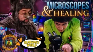
Microscopes & Healing
Episode Discussion
Share your thoughts on this Beakman's World episode